BACKGROUND:
Humeral avulsion of the glenohumeral ligament (HAGL) lesions are associated with shoulder instability. Arthroscopic repair of anterior HAGL lesions typically requires the placement of an anterior-inferior (5-o’clock) portal, with different variations of this portal described. The purpose of this study was to determine the efficacy of described anterior-inferior shoulder arthroscopy portals for arthroscopic anterior HAGL repair, as well as evaluate the safety of these portals with respect to the surrounding neurovascular structures. Additionally, we sought to evaluate the effect of arm adduction vs. standard abduction during anterior-inferior portal creation.
METHODS:
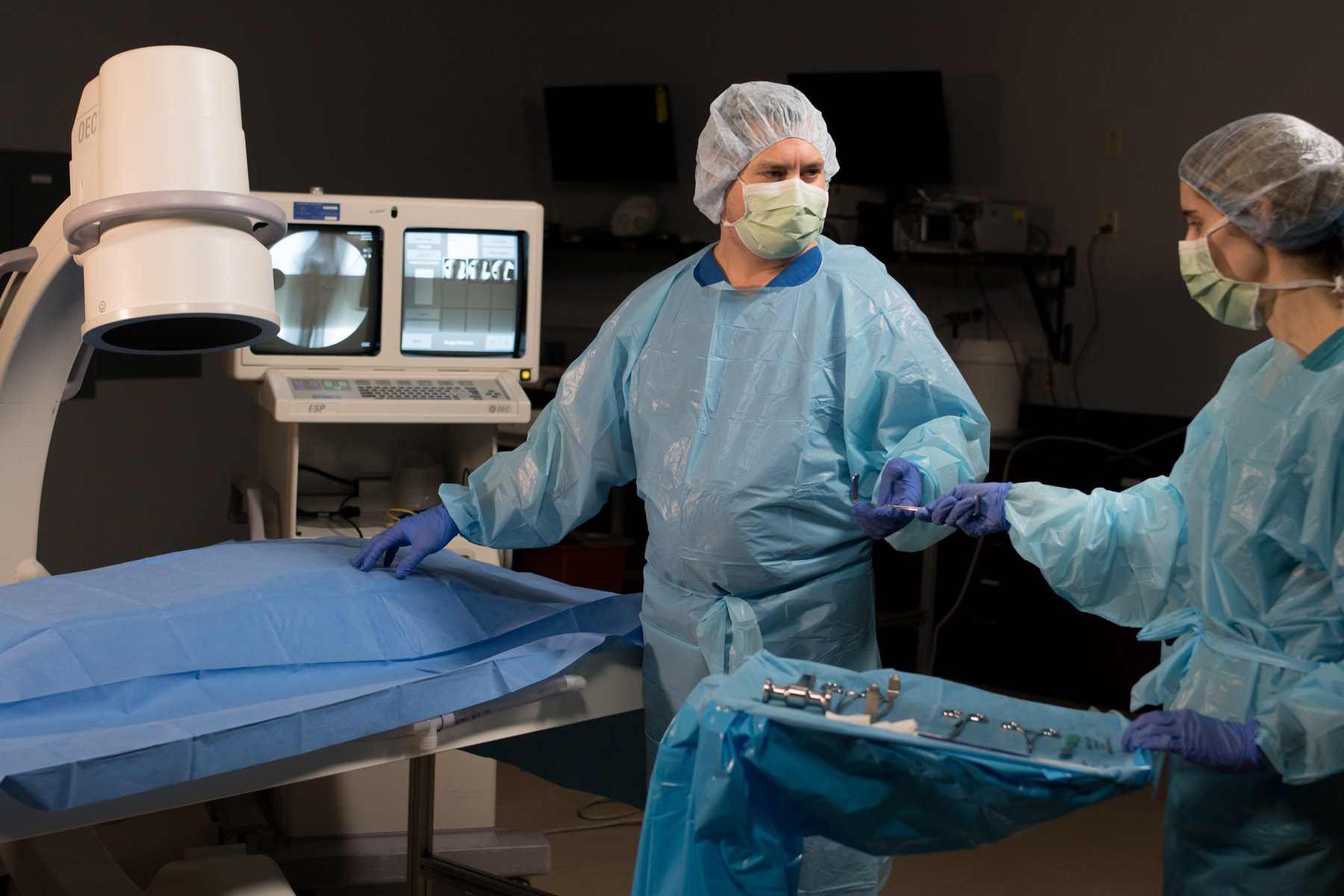
HAGL lesions were created and repaired using an all-arthroscopic technique in 12 cadaveric shoulders (matched pairs). Half of the repairs were performed using a standard 5-o’clock portal, whereas the other half of the matched pairs were repaired using a medialized 5-o’clock portal. Repairs were timed, and the number of anchor pullouts was recorded. The shoulders were subsequently dissected to measure the proximity of the portal to the cephalic vein, musculocutaneous nerve, axillary nerve, and lateral cord of the brachial plexus.
RESULTS:
The average time for HAGL repair was 18.0 ± 4.6 minutes. Repair times using the medial 5-o’clock portal (19.0 ± 3.3 minutes) vs. standard 5-o’clock portal (16.2 ± 5.8 minutes) were not significantly different (P = .37). From abduction to adduction, the cephalic vein distance from the standard 5-o’clock portal increased from 4.1 ± 4.7 mm to 5.2 ± 5.4 mm (P = .02); musculocutaneous nerve distance, from 14.4 ± 9.8 mm to 18.1 ± 10.8 mm (P = .005); axillary nerve distance, from 19.2 ± 9.6 mm to 19.8 ± 9.2 mm (P = .12); and distance of the lateral cord of the brachial plexus, 13.8 ± 6.6 mm to 16.7 ± 6.4 mm (P = .0006).
CONCLUSION:
The arm abduction angle significantly affects the distance of the cephalic vein, musculocutaneous nerve, and lateral cord of the brachial plexus from the anterior-inferior portal, regardless of which portal—standard or medial 5-o’clock portal—is chosen. This portal should be created with the arm in adduction. Arthroscopic HAGL repair can be performed safely, although accurate anchor placement remains a challenge. There was no advantage to use of the medial 5-o’clock portal. With a curved guide, the standard 5-o’clock portal allows for reproducible anchor placement and is recommended for anterior HAGL repairs.